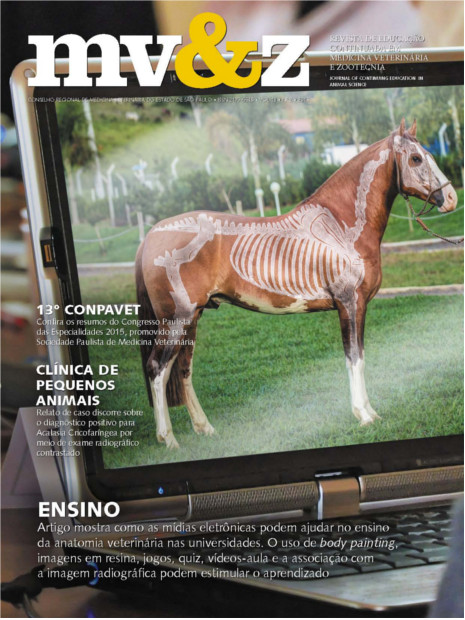
Diagnóstico radiográfico de Acalasia Cricofaríngea em cão – Relato de Caso
Barra lateral de artigos
Conteúdo do artigo principal
Resumo
A acalasia cricofaríngea é uma doença caracterizada pela interrupção da passagem dos alimentos através do esfíncter esofágico cranial, condição essa associada com a incapacidade do relaxamento muscular durante a deglutição cricofaríngea ou com as descoordenadas contrações dos músculos da faringe. Um cão, fêmea, Golden Retriever, seis meses de idade, que apresentava regurgitação, foi avaliado. Na radiografia contrastada do esôfago cervical foi visibilizada a presença do meio de contraste na laringe e no esôfago, no momento da deglutição, e quantidade discreta de bário na traqueia proximal. Após exame radiográfico, o animal foi submetido a ato operatório sendo constatada melhora no quadro clínico. Embora o melhor método de avaliação por imagem seja a fluoroscopia, o objetivo do presente relato de caso é o de discorrer sobre o diagnóstico positivo para acalasia cricofaríngea mediante exame radiográfico contrastado.
Detalhes do artigo
Seção
1. Autores mantém os direitos autorais e concedem à revista o direito de primeira publicação, com o trabalho licenciado sob a Creative Commons Atribuição-NãoComercial-SemDerivações 4.0 Internacional
2. Autores têm autorização para assumir contratos adicionais separadamente, para distribuição não-exclusica da versão do trabalho publicada nesta revista (ex.: publicar em repositório institucional ou como capítulo de livro), com reconhecimento de autoria e publicação inicial nesta revista.
3. Autores têm permissão e são estimulados a publicar e distribuir seu trabalho online (ex.: em repositórios instituicionais ou na sua página pessoal) a qualquer ponto antes ou durante o processo editorial, já que isso pode gerar alterações produtivas, bem como aumentar o impacto e a citação do trabalho publicado (Veja O Efeito do Acesso Livre);